


Bronchitis (bronchitis; bronch[i] + -itis) is an inflammation of the bronchi. This term refers to both separate nosological forms - acute bronchitis and chronic bronchitis. Bronchitis, and secondary bronchial inflammation that develops in the course of a number of diseases, in particular chronic pneumonia, bronchial tumors, heart disease (stagnant bronchitis), kidney disease (uremic bronchitis), etc. In case of catarrhal inflammation the lesion is limited by bronchial mucosa (endobronchitis), at purulent, more severe bronchitis the middle or all bronchial layers are involved (meso- and panbronchitis); at destructive process the inflammation can transfer to peribronchial and interstitial lung tissue (peribronchitis, interstitial peribronchial pneumonia). The role of B. in respiratory pathology is not limited to bronchial tree lesions. According to B.E. Votchal, K.G. Nikulin, A.I. Strukov, I.M. Kodolova and others, it is often the main pathogenetic link in the development of pulmonary emphysema, chronic pneumonia, bronchiectasis, pneumosclerosis.
Classification
There is no generally accepted classification of bronchitis. According to the etiology, a distinction is made between bronchitis viral, bacterial, caused by physical factors (dry air, cold air, etc.), chemotoxic (from inhalation of chlorine fumes, nitrogen oxides, sulphur dioxide, exhaust gases, etc.), dust-related. The type of inflammation, which can be judged by the quality of the sputum, is divided into catarrhal, purulent, putrid, hemorrhagic, fibrinous and mixed, and by the extent of inflammation - into limited and diffuse B.
According to the course of B. it is divided into acute and chronic; depending on the functional disorders there are nonobstructive and obstructive forms of B., and according to the presence of complications there are B. which are uncomplicated and complicated by asthmoid syndrome, peribronchitis (peribronchial pneumonia), lung emphysema, etc.
Statistics
B. is a common disease and one of the most frequent diseases of the respiratory system. Morbidity (according to treatment rate per 1000 people) of adult urban population averages 8.4 for acute B., 10.9 for chronic, with additional registered 4.3 cases of pneumosclerosis and pulmonary emphysema, often combined with B.

The incidence of B. is higher in men than in women. At the age under 40 years old acute B. is registered more often; chronic B. is registered more often at the elder age. B. is more often observed in the older age group. Per 1000 adults of both sexes the incidence figures for acute B. are: at 16-19 years old - 5.0, 20-29 years old - 6.8, 30-39 years old - 10.4, 40-49 years old - 10.3, 50-59 years old - 10.4, 60 years old and over - 7.0, and for chronic B. of both sexes: at 20-29 years old - 7.0, 40-49 years old - 10.2, 60 years old - 10.5. B. respectively: 1.9; 4.4; 8.1; 14.6; 22.5; 23.8 (I. D. Bogatyrev, 1967). I. B. Kalinina (1968) in a mass examination of persons aged 10 years and older has revealed chronic B.D. in 8.6 % of cases. B. in 8.6% of cases. According to Oswald and Medvei (N. C. Oswald, V. C. Medvei, 1955), the incidence of B. is higher in England, and even among office workers the incidence was 13%. B. accounts for 1/4-1/3 of all temporary disability from diseases of the bronchopulmonary system (I. P. Zamotaev, 1969). According to I. D. Bogatyrev (1967), 2.3% of acute and 5.7% of chronically ill patients are hospitalized. B. are hospitalized.
Etiology
Viral and bacterial infections play a major role in the occurrence of B.: influenza, parainfluenza, whooping cough, measles, ornithosis, as well as staphylococcal, streptococcal, pneumococcal and other infectious lesions of the airways. The development of B. is promoted or directly caused by various adverse physical effects - cooling, inhalation of dry air, irritating dust. Less often the main etiolic factor is the effect of toxic chemicals (acid and alkaline vapours, nitrogen oxides, exhaust gases) on the bronchial walls, including injuries caused by poisonous substances (chlorine, phosgene etc.), as well as intoxications during pathological processes (uremia). The course of B. under the influence of physical or chemotoxic factors, as a rule, is complicated by bacterial inflammation of the affected bronchial walls; in such cases we can speak of a mixed etiology of B.
Pathogenesis
In acute B. two phases of development are distinguished - neuroreflexive and infectious. Under influence of cooling, physical and chemical factors or infection conditions for pathological vascular reflexes, metabolic and trophic disorders in bronchial walls are created, as a result of which active hyperemia of mucous membrane with subsequent venous stasis in bronchi develops, mucus secretion increases, barrier role of ciliated epithelium decreases, motor and evacuatory functions of bronchi are violated. All this leads to decrease of barrier function of mucous membrane in relation to bacteria populating it and to decrease of drainage function of bronchi, which promotes development of infectious process. In case of significantly reduced resistance of the body or increased virulence of microorganisms and viruses, the inflammatory process spreads along the length of the bronchus and to all layers of its walls. It is not always possible to distinguish between reflex and infectious phases of bronchitis. Their expression is individual: in some cases reflex reactions prevail, in others, on the contrary, infectious ones. The functional state of the nervous system, which determines neuroreflex reactions, is of great importance in the genesis of B.
Among the factors contributing to the transition of acute B. to chronic, of particular importance are:
- a decrease or change in the reactivity of the macro-organism as a result of lack of hardening, debilitating diseases, repeated cooling, allergic predisposition, etc.;
- irritation of the bronchi by nonspecific irritants (smoking, inhalation of cold or dusty air, the effect of alcohol);
- profound disorders of neurohumoral regulation and trophism of the bronchi in the acute phase of B., leading to restructuring of the bronchial epithelium with quantitative and qualitative disorders of mucus secretion;
- disorders of bronchial drainage function due to damage to the ciliated epithelium and bronchial motility disorders, as well as due to bronchial obstruction, especially in inflammatory and (or) allergic bronchospasm;
- the presence of chronic foci of infection in the upper respiratory tract, especially in the sinuses.
Acute bronchitis with bronchial permeability disorders have the highest probability of transition to chronic course (B. E. V otchal, 1967; J. N. Dotsenko, 1958, and others). Apparently, the frequent combination of obstructive B. with immunity disorders has significance.
Experimentally, in animals, chronic B. B. are caused in experimental animals by tracheotomy with introduction of infected foreign material. To disturb bronchial drainage and bronchial patency, the tracheal lumen is reduced with a plastic ring. Cooling or administration of alcohol is used to lower the body's resistance (V. V. Dubelay, 1963). Ε. M. Altman (1968) experimentally obtained a model of inflammation of bronchi and lungs by infecting the palatine tonsils. Hist. preparations revealed inflammatory infiltrates in submucosal and mucosal layers of bronchial wall, mucous membrane was sloughed off in places, there was accumulation of leukocytes in bronchial lumen.
Acute bronchitis
Most frequently acute bronchitis develops as the result of viral or bacterial infection against a background of cooling, less often - against irritating influence of physical and chemical factors.
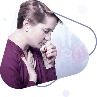
The clinical picture of acute IB consists of symptoms of general intoxication and symptoms of bronchial involvement. Body temperature rises in the first 2-3 days, but often remains normal. There is general weakness, sometimes coughing, muscle pain in the back and extremities, runny nose, hoarseness of voice, tickling in the throat. The cough at first is dry, rough, with scanty viscous sputum. On the 2nd or 3rd day of the disease, there are burning sensations behind the sternum, which intensify when coughing. As the process spreads along the course of the bronchi, the symptoms of upper respiratory tract irritation weaken, and the process seems to move downward, the cough comes from deep inside, spitting up becomes lighter, the sputum is excreted in larger amounts, it becomes mucopurulent. Percussion sound over the lungs is unchanged, auscultation reveals rigid vesicular breathing and depending on the nature of the sputum (liquid or viscous), silent wet or dry, usually scattered rales are heard. With viscous secretion in the large and medium-sized bronchi, the rales are low, buzzing (rhonchi sonores), with secretion in the small bronchi or with swelling of their mucous membranes, the rales are high, whistling (rhonchi sibilantes).
A number of features of clinical symptomatology of acute bronchitis are determined by the state of external respiratory function and bronchial patency disorders (obstructive and nonobstructive bronchitis). In obstructive bronchitis small bronchi are affected. Bronchial patency disorders are caused by increased tone of bronchial muscles, mucous membrane swelling and mucus overproduction. The specific significance of these factors in patients varies, but the leading role in the mechanisms of bronchial patency impairment is played by neuroreflex factors manifested by bronchospasm. Reflexes can come from irritation of bronchial interreceptors and upper airways by pathological process. Swelling of the mucous membrane depends on the degree of its hyperemia and severity of inflammatory edema. Retention of secretion depends on its viscosity. A patient with obstructive B may feel dyspnea at usual physical exertion, sometimes even at rest; there is various degrees of lengthening of exhalation phase, when percussing the chest there is some tympanic sound, breathing is hard vesicular, hissing, more persistent on exhalation. Sometimes you have to identify them by listening to a patient standing or lying down, when exhaling forcibly. Patients of this group often have coughing fits, which is followed by shortness of breath for some time. Recovery of bronchial patency in acute B. is observed in different terms. From instrumental investigations reliably and with great completeness the bronchial permeability disorders are revealed by pneumotachometry and the study of forced vital capacity spirographically.
Acute B in elderly people with involvement of small bronchi has a severe course. Due to bronchial permeability disorders and senile emphysema, breathing becomes frequent and superficial, dyspnea and diffuse cyanosis occur. On the CNS side, at first there is anxiety, agitation, which later turns into apathy and somnolence. Heart tones are muffled, pulse is rapid. Respiratory failure may join cardiac.
The course of acute B., especially when small bronchi are affected, can be complicated by pneumonia, both at the expense of atelectasis infection, and due to the transition of inflammation to the interstitial tissue of the lung. The patient's general condition worsens, with chills, fever, increased cough, pus-like sputum, and dyspnea may occur. Complications of small focal pneumonia are especially frequent in the elderly and the elderly. Percussion sound over the lungs becomes shortened or with tympanic tinge, breathing is rigid vesicular, localized moist small vesicular rales are heard, bronchophony is often amplified. The blood shows neutrophilic leukocytosis, RPE is accelerated.

Diagnosing of acute B. does not cause difficulties and is established taking into account etiological factor by leading signs: the most important of them are cough, separation of sputum and listening to dry and (or) moist rales in lungs against hard breathing.
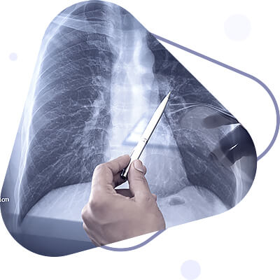
X-ray diagnosis of acute B. is limited to recognition of functional disorders associated with impaired ventilation of the bronchi due to their spasm, swelling of the mucous membrane and retention of bronchial secretion. On routine radiographs and electroradiographs against the background of general pulmonary bloating focal or lamellar atelectasis may be seen, and sometimes small areas of pneumonia, complicating acute B. Respiratory mobility of the diaphragm is limited.
The prognosis in acute B., as a rule, is favorable. In most cases, especially in the catarrhal form, the disease ends with recovery with restoration of the normal condition of the bronchial walls and lumen. In some cases, especially with bronchial permeability disorders, the acute process becomes chronic. In cases of purulent B. after recovery fibrous thickening of the bronchial wall may remain, often with narrowing of its lumen.
With pronounced and predominant lesion of small bronchi (bronchiolitis) the outcome of acute bronchitis may be overgrowth of the bronchial lumen with connective tissue - obliterating B. A similar outcome is often observed in acute chemotoxic B. (after inhalation of acid vapors, chlorine, phosgene, diphosgene, etc.), as well as in B. against the background of some viral infections (measles, influenza). Temporary disability depends on the degree of bronchial wall damage (in endobronchitis it is brief, in panbronchitis it can last up to several weeks) and on the extent of the damage, which determines the degree of functional disorders (in cold nonobstructive B. the duration of temporary disability usually does not exceed 5-7 days, in obstructive it increases to 2-3 weeks).
Treatment of acute B. should be early, taking into account the etiology and pathogenesis of the disease. In viral and bacterial B., often developing during epidemic respiratory infections (influenza, parainfluenza, etc.), etiotropic therapy, as well as pathogenetic and symptomatic treatment of B itself is carried out.
A patient with acute B. should keep to bed rest and avoid cooling, but stay in a ventilated room with cool, fresh air. It is recommended to drink plenty of warm diapers (tea with raspberry jam, limeblossom tea), take alkaline (Borjomi with milk, drinking soda with milk) from the beginning of the disease, with painful sensations behind the chest - mustard pads on the chest area, interscapular area, circular banks, warming compresses, mustard foot baths. With a dry painful cough, cough suppressants shall be used at the beginning of the disease - codeine, kodoterpine, dionine. Since the intensification of sputum separation and difficult coughing appointment of cough suppressants is contraindicated, during this period, appointed cough suppressants, such as infusion of Thermopsis (0.6 or 1.0 to 200.0), 1 tablespoon every 2-3 hours. In cases of bronchial obstruction bronchodilators are individually selected - ephedrine, atropine, preparations of belladonna, theophedrine, antastman, eufillin in suppositories.
With purulent sputum sulfonamides or antibiotics are indicated; the latter are rationally prescribed in the form of aerosols 2-3 times a day. In cases of bronchobronchiolitis, antibiotic or sulfonamide therapy is combined with (for adults) 30-40 mg daily of prednisolone (or equivalent doses of triamcinolone, dexamethasone) for 5-7 days, usually until the high-pitched dry rales in the lungs have disappeared. For this duration of use, hormones may be withdrawn immediately, but in cases of a longer course of therapy, they are withdrawn gradually. Cardiovascular agents are indicated in the presence of heart lesions, especially in the elderly. In these cases, oxygen therapy is very effective.
In order to restore the impaired blood circulation in the mucous membrane of the bronchi, trachea and nasopharynx in catarrhal B., if tuberculosis is excluded, quartz irradiation of the surface of the chest with one biodose daily with an area of 400-600 cm2 is prescribed. Diathermy of thoracic area or inductotherapy on interscapular area is reasonable in more profound B.
Prevention of acute B. consists of hardening the body, compliance with hygienic rules at work and at home, carrying out anti-flu vaccinations. Timely and persistent treatment of upper respiratory infections is important:
- rhinitis;
- sinusitis;
- tonsillitis;
- pharyngitis.
A person with B. should be isolated at home. People in contact with B. should wear masks.
Chronic bronchitis
The first clinical description of chronic bronchitis is by R. B. is by R. Laennec (1826). G.I. Sokolsky in 1839 pointed out the connection of all chronic inflammatory lung diseases with bronchitis. A.N. Rubel (1925) has considered that the basis of chronic bronchitis is pneumosclerosis. pneumosclerosis is the basis of chronic bronchitis.
Despite its apparent simplicity, reliable diagnosis of chronic bronchitis is difficult and always requires a diagnosis of chronic bronchitis. It is difficult to make a reliable diagnosis of chronic bronchitis and it is always necessary to distinguish it from chronic lung diseases (tuberculosis, cancer, bronchiectasis, dry pleurisy, bronchial asthma, mycosis etc.). In a number of countries, chronic B. B. are considered to be the most frequent pathology of the respiratory system. It should be noted, however, that, for example, most English researchers under the term "chronic bronchitis" understand not only B. proper, but also complicated forms of the disease, including chronic pneumonia, bronchiectatic changes, emphysema of lungs, pulmonary heart.
The WHO Expert Commission guides in the diagnosis of chronic B. B. on the presence of cough and sputum secretion for many days or up to 3 months in each of the next two years, unless associated with localized bronchopulmonary disease. These symptoms are not exhaustive, as up to 20% of patients with chronic B. B. do not have any complaints, and B. is diagnosed by the detection of wheezing rales in the lungs and decreased ventilatory function.
The decisive role in the etiology of chronic B. The decisive role in chronic B. etiology is played by recurrent upper respiratory tract catarrh, relapses of acute B. and long-term influence of physical and chemical damaging factors. Chronic B. B. can develop as a result of acute B. Under the influence of long-term harmful factors, it often develops chronically primary bronchitis. In the pathogenesis and course of chronic bronchitis diseases of nasal. Diseases of the nose, nasopharynx, and sinuses are particularly important in the pathogenesis and course of chronic bronchitis. Foci of infection contribute to reinfection of the bronchi. In addition, impaired air purification and warming functions and disabling of the naso-pulmonary reflex involved in the regulation of breathing depth should be taken into account.
Chronic bronchitis and its exacerbations are promoted by repeated cooling. The maintenance of chronic bronchitis and its exacerbations is promoted by repeated cooling, chronic bronchial irritation by tobacco smoke in smokers, inhaled dust, harmful gases. It seems that immune disorders, in particular autoallergies, play an important, but poorly studied role.
Pathological anatomy. Changes of bronchial wall in chronic B. B. are various and depend on character of inflammation, affection depth, bronchial caliber, reason, causing B, and disease duration.
According to the distribution of chronic bronchitis. It can be diffuse and limited (lobes, segments). At limited processes, changes are more often observed in those segments, where bronchopneumonic foci arise with greater constancy (6th, 9th, 10th segments of lower lobes, 1-2th in upper lobes, and 4th-5th segments of left lung).
Catarrhal B is characterized by loose infiltration of mucous membrane with histiocytes, lymphoid, plasma cells with leukocytes admixture, its full bloodiness and edema, mucus hypersecretion by covering epithelium and mucous glands, epithelium sloughing. Epithelium metaplasia into multilayer squamous epithelium, enlargement of bocal cells (replacement of ciliated cells by bocal cells), enlargement of mucous glands and exit ducts, accumulation of SHIK-positive secretion with γ-metachromasia intensification in them occurs.
In purulent B. cellular infiltrates with admixture of leukocytes diffusely permeate mucous membrane and spread to submucous layer. The basal membrane is thickened, the epithelium shows hypersecretion, metaplasia in places. There may be ulceration of the mucosa. There is purulent exudate in the bronchial lumen. In large (segmental) bronchi, muscle bundles, elastic fibers, mucous glands, cartilaginous plates remain unchanged for a long time.
In destructive B the mucous membrane, all layers of bronchial wall and peribronchial tissue are diffusely infiltrated by histiocytes, lymphoid and plasma cells. In some places cellular infiltration turns into granulation tissue, replacing the bronchial wall, subsequently undergoing sclerosis. The mucosa is thickened, and forms polypoid outgrowths, consisting of a base infiltrated by cells and granulation tissue - destructive-polyposis B.; there may be ulceration of the mucosa - destructive-ulcerative B.

In submucosal layer and mucosa among infiltrate cells and in lymphoid follicles, formed in large numbers in peribronchial tissue, there is an accumulation of plasma cells with pyroninophilic granularity and with SHIK-positive material in cytoplasm. The basal membrane is thickened, sclerosed. SHIK reaction reveals varying degrees of bronchial epithelium and mucosal glands secretion, desquamation of epithelium, focal metaplasia of epithelium into multilayer squamous epithelium are observed. Infiltrative and sclerotic changes in submucosal layer are combined with more severe mucosal changes, desquamation of epithelium, sclerosis of stroma and basal membrane, atrophy of glands and their hyposesecretion, cystic expansion of glands. As a result of inflammatory infiltration and development of granulation tissue there is gradual disintegration of muscle bundles and elastic fibers, their uneven distribution in the bronchial ring and gradual atrophy. Fatty dystrophy develops in muscle bundles, glycogen content decreases. Cartilage plates undergo dystrophic changes, in which one can see resorption of the main substance of cartilage by granulation tissue, calcification, development of bone and bone marrow tissue.
Nerve fibers and intramural ganglia of bronchial walls develop differently pronounced dystrophic changes characterized by varicose fibers, homogenization, vacuolization and granular decay of axial cylinders, vacuolization and shriveling of nerve cells. Bronchial arteries develop productive vasculitis with sclerosis, obliteration of lymph vessels.
Infiltrative-productive and destructive changes of structural and functional elements of bronchial wall (muscle bundles, elastic fibers, cartilage plates, nerve elements) are most pronounced in small bronchi (subsegmental and smaller caliber). As a result there occurs gradual dilatation of lumen and deformation of bronchial walls with development of bronchiectasis.
The clinical picture of chron. Bronchitis is determined by the caliber of affected bronchi (affecting large, medium, small bronchi), its extent (segmental, diffuse bronchitis) and the depth of bronchial wall damage. The main symptoms of chronic. Б.:
- cough, sometimes of a seizure-like character, more often in the morning after sleep or when the temperature of inhaled air changes;
- separation of mucopurulent sputum, less often of purulent nature;
- general malaise;
- in obstructive B. - shortness of breath at physical load, intensity of which varies during the day due to variations of atmospheric pressure, temperature of exhaled air and its humidity, degree of sputum accumulation and coughing up.
When the large bronchi are affected, the cough is periodic or episodic, the sputum is mucopurulent or purulent, the amount is moderate, sometimes very scanty. Percussive and auscultatory findings without deviation from normal. When the process is localized in the middle bronchi, the cough is moist, more often in the morning after sleep, with mucopurulent sputum. Percussion sound is unchanged or may have a boxy tint; auscultatively, dry bass rales are detected, which easily disappear when coughing. If the process is localized in the small bronchi, in addition to cough, sputum secretion, shortness of breath is noted. The amount of sputum is small, and the cough is moderate. Percussion sound with boxy tinge, auscultatively dry high-pitched (whistling) rales prevail. Examination of the external respiratory function by pneumotachometry or Votchal-Tiffno test reveals the presence of bronchial obstruction. The mechanisms of bronchial obstruction are varied: in addition to bronchospasm, edema and swelling of bronchial mucosa, hypersecretion, inflammatory infiltration of the bronchi are important. The leading factor is bronchospasm, in the pathogenesis of which the excessive production of bronchospastic substances (histamine, serotonin, bradykinin) during immunological antigen-antibody reaction or oversensitivity of bronchial tree receptors, overreacting to normal physical or chemical stimuli due to inflammation, is especially common. Increased sensitivity of bronchial receptors is especially pronounced after influenza, but in patients with chronic B. B. it is almost constant. When differentiating between allergic and inflammatory nature of bronchospasm it is necessary to consider allergic predisposition, which in patients with diffuse form of peribronchial chronic pneumonia with asthmatic B. is found in 76.8% of cases.
In the course of chronic. B can be distinguished between remission and exacerbation phases. In remission the main clinical manifestations are expressed moderately (rare cough with little sputum, dyspnea without a tendency to progression) or only bronchial permeability disorders are revealed instrumentally. At exacerbations of chronic. B., which are often seasonal, the severity of clinical manifestations increases.
It should be kept in mind that except for periods of acute respiratory infection (viral or bacterial), there are no symptoms of general intoxication in chronic bronchitis. B. are absent. If subfebrile fever and signs of general intoxication (sweating, malaise, weakness, etc.) are detected, it is necessary to think about peribronchitis - process transfer to peribronchial interstitium, i.e. interstitial pneumonia.
Bronchial dysfunction is manifested as bronchospasm (local or generalized), dyskinesia (violation of the complex of normal bronchial movements during breathing), as well as delayed evacuation in the phase of disease exacerbation.
Complications. In chronic. With total bronchial wall lesions conditions are created for infection spreading, especially from small bronchi, to interstitial tissue of lungs, which leads to development of interstitial pneumonia and pneumosclerosis. The development of pneumonia in chronic B. Local ventilation disorders often contribute to the development of chronic pneumonia: blockage of bronchial lumen by secretion or swollen mucous membrane leads to the formation of lung atelectasis, complicated by bronchopneumonia. Acute interstitial or parenchymatous pneumonia in case of insufficiently vigorous treatment is prone to transition to chronic, which contributes to the same violations of ventilation and bronchial drainage. Therefore, it is important to diagnose pneumonia early, with initial symptoms. These include subfebrile fever, coughing, sweating of the upper half of the trunk, increased purulent sputum, blood reactions (neutrophilic shift, accelerated RPE).
Chronic obstructive bronchitis is followed early by pulmonary emphysema, diffuse pneumosclerosis and chronic pulmonary heart develop. In atrophic processes in bronchial wall or in local peribronchial pneumosclerosis, bronchiectases are formed.
The diagnosis of chronic. Bronchitis B. is based on the history (frequent colds in a patient), clinical symptoms (cough, sputum discharge), hearing rales in the lungs, and functional test data (decreased expiratory pneumotachometry). Among instrumental methods, bronchography and bronchoscopy are valuable.
Radiological examination in chronic bronchitis. It allows to reveal the affected area and determine the degree and nature of pathological changes in bronchi and lung tissue. The latter are detected on radiographs only when inflammatory process is not limited to endobronchitis, and leads to pan- and peribronchitis with development of interstitial tissue infiltration.
Radiomorphological and roentgenofunctional picture of lesions in chron. B. is studied by fluoroscopy, radiography or electroradiography, bronchography or bronchokinematography.
Four groups of X-ray symptoms can be distinguished:
- changes in pulmonary pattern;
- emphysema combined with areas of lobular and acinic thickening;
- morphological changes of bronchi;
- changes in bronchial function.
Changes in pulmonary pattern are expressed in its enhancement due to thickening of bronchial walls, axial sections of which are clearly visible in their orthograde course. In the late stages, with pulmonary hypertension, the pulmonary pattern is intensified due to the vascular component.
Areas of lobular edema, small atelectasis, widespread emphysema are usually observed in acute stage with bronchial permeability impairment.
Morphological symptoms of lesion of large and medium-sized bronchi consist in changes of bronchial contours [irregularity, diverticular-like protrusions], their caliber (alternation of dilated and narrowed sections) and direction (decrease of divergence angles). Small bronchi and bronchioles either are not filled with contrast agent, or changes of bronchioloectasis type are found in them.
The prognosis in chronic B. B. is favorable for life, but unfavorable in terms of complete and persistent cure. The probability of cure of chronic B. The more bronchial permeability disorders are expressed, the less is the probability of chronic B. cure. The prognosis of patients' ability to work and further course of the disease is determined mainly by failure of respiratory function, the rate of development of which depends on the severity of bronchial permeability disturbances, the degree of development of pneumo-sclerosis, emphysema of lungs, complications of chronic bronchial pneumonia. The prognosis worsens considerably in case of development of pulmonary heart, especially in its decompensation stage.
Treatment in chronic B. B. is determined by the stage of the process. In the acute stage the patients need to be released from work until the process completely subsides; they are prescribed bed rest, antibacterial therapy with sulfonamides or antibiotics, plenty of warm drink, expectorants and bronchodilators, as in acute B. Acute bacterial infection should be eliminated as soon as possible, so the primary antibacterial treatment is given without determining the sensitivity of microorganisms to antibiotics.
When prescribing antibacterials, it should be based on the fact that the most frequent exacerbation of chronic bacterial infections is caused by bacilli. B. is caused by Pfeiffer's bacillus, pneumococcus, and staphylococcus aureus. Oxitetracycline 2.0 g per day is prescribed, and if there is no effect on the 5th-7th day - levomycetin 2.0 g per day or ampicillin 2.0-3.0 g per day. In many cases, prolonged sulfonamides are effective, especially in combination with folic acid antagonist (trimethoprim), such as biseptol and others. Usually it is enough to give the antibiotic for 5-7 days, in severe cases 3-4 weeks or longer. To identify the causative agent of the exacerbation and determine its sensitivity to antibiotics, sputum culture for flora should be carried out only by Mulder (J. Mulder, born 1901). In cases of a persistent course, intramuscular antibiotic injection in combination with aerosol inhalation has an effect.
At severe bronchial obstruction with presence of eosinophilia in blood and sputum of patients in complex therapy of chronic. B. are used glucocorticoids.
When the process goes into remission stage, the patients' regimen is determined by the degree of respiratory insufficiency and circulatory disturbances. At chronic obstructive B. the work of respiratory muscles may increase by 5-10 and even 20 times; physical labor is contraindicated for these patients from the early stages of respiratory failure. Providing bronchial drainage is of great importance. In the presence of thick, difficult to separate sputum is recommended to use 3% solution of potassium iodide 1 tablespoon every 2-3 hours for 5-7 days, thermopsis. The expectorant effect of iodine is higher than that of thermopsis. In some cases of obstructive B. there is increased mucus secretion of increased viscosity, to facilitate the separation of which requires the appointment of mucolytic drugs (chymoppsin, chymotrypsin). Effective bronchial drainage is supported by bronchodilators, of which the most reasonable is eufillin, the best is intravenous dropwise 0.24-0.48 g in 5% glucose solution or suppositories 0.4-0.5 g two or three times a day. Of bronchodilators of sympathomimetic type, ephedrine, novodrine (euspiran), alupent are prescribed. Ephedrine acts from 4 to 8 hours, but the effective dose varies individually from 10 to 100 mg. Alupent has a good bronchodilator effect lasting 2-4 hours. It is usually rational to prescribe 1/2-1/4 tablets of Alupent (10 mg) 3-6 times a day at regular intervals. Inhalation of alupent, novodrin is best used in cases of severe obstruction. Inhalation of atropine (0.3-0.5 ml of 0.1% solution) is effective in some cases.
In the absence of bronchodilator effect of atropine, eufillin and presence of contraindications to sympathomimetics, glucocorticoids shall be prescribed with selection of individual maintenance dose.
Patients with chron. Recommended physiotherapy treatment - diathermy, ionophoresis, quartz on the chest area. Complex breathing exercises and postural drainage (rational positioning of the body) should be conducted 2-3 times a day. Indicates climate therapy - in a pine forest, in the steppe zone, in a mountain climate, the resorts of the southern coast of Crimea, in Kislovodsk. In the last phases of chronic B. B. treatment is determined by the condition of respiratory and circulatory systems.
The prophylaxis of chronic Bla. CD, as well as acute CD, consists of individual and general health-improving and hygienic measures. Individual measures:
- prevention of cooling;
- regular hardening of the body;
- exercise and sports;
- smoking cessation;
- sanitation of the upper respiratory tract.
The role of general health and hygienic measures in the home and at work is great, and above all the fight against dust and air pollution by gases and other harmful substances. When tempering necessary to observe the systematic and regularity of the activities. Start tempering with taking air baths and dry body rubs, washing feet before bedtime with water t ° 32-34 ° with a subsequent decrease in temperature after 2-3 days at Ie to tap water temperature.
After getting used to the air baths move to wiping: first, with a wet wrung towel, then a wet not wrung towel, then proceed to poured water indifferent temperature (30-32 degrees) and a subsequent decrease in its temperature by 1 degree every 2-3 days to the tap water temperature. It takes 7-14 days to switch from one cooling intensity to another.
Hardening is inseparable from compliance with the rules of personal hygiene. It is necessary to make sure that you always have dry, seasonally appropriate clothing. Ventilation of rooms, dust control, and preventive measures against the infections that most often cause the development of B., especially against influenza are of great importance.
Specific prophylaxis against viral influenza by active immunization of population with live influenza vaccine and nasal injection of interferon is effective. The early detection and thorough treatment of rhinitis, tonsillitis, inflammation of the accessory sinuses are of great importance in the primary prophylaxis of B.
In each case of chronic B. It is necessary to consult an otorhinolaryngologist to determine the efficiency of nasal breathing, to detect possible infectious foci, and to treat them. Because of the large role of allergies in chron. B. it is necessary to use all means of allergic diagnostics in order to exclude patients from further contact with allergens and perform desensitization. Among the measures of secondary prophylaxis in chron. Constant performance of respiratory therapeutic exercises, procedures of positional drainage of bronchi are of great importance. Prof. measures for chron. B. is also to fight industrial dust and air gasses.
Prevention of progression of respiratory and cardiac failure in chronic bronchitis is largely defined by timely prevention of bronchial hypertension. B is largely determined by the timely and proper employment of patients. Significant physical strain, unfavorable meteorological conditions, influence of dust and airway irritating gases, influence of bronchotropic and pulmotropic poisons, contact with allergens is contraindicated in case of exo-allergies.
Bronchitis in children
Isolated defeat of the bronchial tree in children, unlike adults, is rare.
Acute bronchitis usually accompanies seasonal and epidemic acute respiratory diseases, or appears at the beginning of some infections (measles, whooping cough, etc.). The causative agents of acute B. are various viruses (influenza viruses, adenoviruses, respiratory syncytial viruses, etc.) and bacteria - staphylococci, streptococci, pneumococci, less frequently E. coli, fungi, etc. Non-microbial etiology, e.g. from inhalation of irritants, is rare in children.
Acute bronchitis is most common in young children suffering from exudative-catarrhal diathesis, and in older children with pathology of ENT-organs (rhinitis, adenoiditis, sinusitis, chronic tonsillitis). In this case, the inflammatory process may begin in the upper respiratory tract with subsequent spread to the trachea and bronchi.
Clinical picture
Along with irritability, lethargy is observed; the temperature usually does not rise significantly and fluctuates around subfebrile numbers for several days. Preschool and school-age children sometimes complain of pain behind the chest and headache. A characteristic symptom of acute B is a dry cough at first, painful, more often in the evening and at night, and after 1-2 days it becomes wet, with mucopurulent or purulent sputum, which children usually swallow. On examination of the lungs, there may be a boxy percussion sound. Exhalation is prolonged; as a rule, scattered rales are heard, at first dry and then wet coarse and medium bubbling.
The inflammatory process is not limited to the bronchial mucosa; usually all layers of the bronchial wall are involved, and in some cases peribronchial tissue as well. Swelling of the bronchial wall, presence of mucus and sloughing epithelium in the bronchial lumen impair bronchial patency.
In young children in some cases atelectasis of pulmonary tissue occurs, contributing to the development of pneumonia (Η. F. Filatov, Yu. F. Dombrovskaya).
Radiological picture
At acute B. some dilation and thickening of shadows of lung roots and strengthening of lung pattern shall be noted.
A characteristic picture in patients with B. can be observed during bronchoscopy:
- edema;
- hyperemia;
- readily occurring bleeding of the mucous membrane;
- mucopurulent contents in the bronchial lumen.
Inflammatory changes are usually diffuse (diffuse B.); more pronounced bronchial changes in separate segments are possible. In some cases (with foreign bodies, abnormal development of airways, etc.) local changes of lung segments or lobes are observed (local B). In smears and secretions obtained from bronchi, microscopic examination reveals leukocytes, cells of bronchial mucosa and mucous glands, and in allergic conditions - a significant number of eosinophils. In some cases B. can be accompanied by asthmatic syndrome, requiring differential diagnosis with bronchial asthma in children.
At the protracted and recurrent course of acute B. development of chronic B. which is usually combined with bronchial asthma is possible. It is usually combined with chronic lesions of interstitial tissue and lung parenchyma, i.e. with chronic nonspecific pneumonia.
Differential diagnosis of acute bronchitis B should be done with pneumonia, pulmonary tuberculosis; some infectious diseases (measles, whooping cough, flu, etc.) are excluded depending on the clinical picture as well as epidemiological situation.
The prognosis for acute B. is favourable. It is possible that the process will spread and bronchiolitis and pneumonia will develop.
Treatment depends on the severity of the disease. During mild and moderate acute bronchitis patients are prescribed salicylates, expectorants and phlegm thinners, multivitamins, plenty of fluids (juices), steam and alkaline inhalations, hot baths, jars, mustard powder and wet mustard wraps, chest irradiation with sunlux lamp, UVA of collar zone and inter-scapular area; patients with prolonged bronchitis are also recommended inductothermy and microwave therapy. Severe course requires sulfonamides or antibiotics for 5-7 days.
Prevention of acute B consists of hardening procedures, normalization of the daily routine, adequate nutrition, sufficient exposure to air, protection from contact with patients with acute respiratory and infectious diseases, timely treatment of ENT-organ diseases, etc.
By: Dr. Jai Jalaj