Laboratory and instrumental methods of investigation
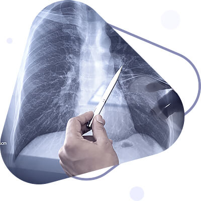
Fluoroscopy, radiography, bronchography, and lung tomography are used to examine the respiratory organs. Fluoroscopy is the most common method of examination, which allows you to visually determine changes in the transparency of lung tissue, detect foci of compaction or cavities in it, to identify the presence of fluid or air in the pleural cavity, as well as other pathological changes. Radiography is used to record and document changes in the respiratory organs detected during fluoroscopy on X-ray film. In pathological processes in the lungs that lead to loss of airiness and thickening of lung tissue (pneumonia, pulmonary infarction, tuberculosis, etc.), the corresponding parts of the lungs on the negative film have a paler image compared to the normal lung tissue. A cavity in the lung containing air and surrounded by an inflammatory roll looks like a dark oval-shaped spot surrounded by a paler shadow than the shadow of the lung tissue on the negative x-ray film. The fluid in the pleural cavity, which transmits fewer X-rays compared to the lung tissue, gives a paler shadow on the negative X-ray film compared to the shadow of the lung tissue.
The radiological method makes it possible to determine not only the amount of fluid in the pleural cavity, but also its nature. If there is inflammatory fluid or exudate in the pleural cavity, its level of contact with the lungs has a oblique line gradually heading upward and laterally from the midclavicular line; if noninflammatory fluid or transudate has accumulated in the pleural cavity, its level is more horizontal. Tomography is a special radiographic technique that allows a layer-by-layer radiographic examination of the lungs. It is used to diagnose bronchial and lung tumors, as well as small infiltrates, cavities, and caverns at different depths of the lungs.
Bronchography is used to examine the bronchi. After a preliminary anesthesia of the respiratory tract, a contrast agent is injected into the lumen of the bronchi that detains X-rays (e.g., idolipol), then the lungs are radiographed and a clear image of the bronchial tree is obtained on the radiograph. This method makes it possible to diagnose bronchial dilatation (bronchiectasis), lung abscesses and caverns, and narrowing of the lumen of the large bronchi by a tumor or a foreign body. Fluorography is also a type of radiographic examination of the lungs. It is performed using a special apparatus, a fluorograph, which allows taking an X-ray picture on small-format film, and is used for mass preventive examination of the population.
Endoscopic examination. Endoscopic methods of examination include bronchoscopy and thoracoscopy. Bronchoscopy is used to examine the mucous membrane of the trachea and bronchi of the first, second and third order. It is performed with a special device - bronchoscope, which is attached to special forceps for biopsy, extraction of foreign bodies, removal of polyps, photo attachment, etc. Before introducing the bronchoscope, anesthesia of 1-3% dicaine solution of the mucosa of the upper respiratory tract is carried out. The bronchoscope is then inserted through the mouth and vocal slit into the trachea. The investigator examines the mucosa of the trachea and bronchi. Using special forceps on a long handle, a piece of tissue can be taken from the suspicious area (biopsy) for histological and cytological examination, and it can also be photographed.
Bronchoscopy is used for diagnosing erosions, ulcers of the bronchial mucosa and tumors of the bronchial wall, extraction of foreign bodies, removal of bronchial polyps, treatment of bronchiectatic disease and centrally located lung abscesses. In these cases, purulent sputum is first sucked through the bronchoscope and then antibiotics are injected into the bronchial lumen or cavity. Tore the manipulation field with iodine and alcohol and local anesthesia at the puncture site. The puncture is usually performed along the posterior axillary line in the seventh or eighth intercostal space along the upper edge of the rib. For diagnostic purposes, 50-150 ml of fluid is taken and sent for cytological and bacteriological examination. For therapeutic purposes with accumulation of large amount of fluid in pleural cavity 800-1200 ml of fluid is initially taken. Removal of more fluid from pleural cavity leads to quick displacement of mediastinal organs to the sick side and may be accompanied by collapse. To extract the fluid a special 50 ml syringe or Poten's apparatus is used. The fluid obtained from the pleural cavity may be of inflammatory (exudate) or noninflammatory (transudate) origin. For the purpose of differential diagnosis of the nature of the fluid its specific gravity, the amount of protein, erythrocytes, leukocytes, mesothelial and atypical cells contained in it are determined. Specific gravity of inflammatory fluid is 1,015 and higher, protein content is more than 2-3%, Rivald's test is positive. The specific gravity of the transudate is less than 1,015, the amount of protein is less than 2%, Rivald's test is negative. The Rivald test involves taking a 200 ml cylinder, filling it with tap water, adding 5-6 drops of strong acetic acid, and then pipetting a few drops of pleural fluid into it. The appearance of a cloudy cloud at the point where the drops dissolve indicates the inflammatory nature of the pleural fluid, which contains increased amounts of serosomucin (positive reaction, or Rivald's test). Non-inflammatory fluid does not give a cloudy cloud (negative Rivald's test).
Sputum examination. Sputum is a pathological secretion of the respiratory organs, ejected when coughing and expectorating (normal bronchial secretion is so insignificant that it is eliminated without expectoration). Sputum may include mucus, serous fluid, blood and respiratory cells, elements of tissue decay, crystals, microorganisms, protozoa, helminths and their eggs (rarely). Examination of sputum helps to establish the nature of the pathological process in the respiratory organs, and in some cases determine its etiology. Sputum for examination is better to take in the morning, fresh, if possible, before meals and after mouthwash. However, to detect Mycobacterium tuberculosis sputum should be collected within 1-2 days if the patient secretes little of it. Saprophytic flora multiplies in stale sputum and destroys the form elements. The daily amount of sputum varies widely, from 1 to 1000 ml and more. Large amounts of sputum are discharged at once, especially when the patient changes position, characteristic of mesenteric bronchiectasis and bronchial fistula formation in pleural empyema.
Examination of sputum begins with its examination (i.e. macroscopic examination) first in a transparent jar and then in a Petri dish, which is placed alternately on a black and white background. The nature of the sputum is noted, meaning its main components discernible to the eye. The color and consistency of sputum depend on the latter. Mucous sputum is usually colorless or slightly whitish, viscous; it is separated, for example, in acute bronchitis. Serous sputum is also colorless, liquid, frothy; seen with pulmonary edema. Mucopurulent sputum is yellow or greenish, viscous; it is formed in chronic bronchitis, tuberculosis, etc. Purely purulent, homogeneous, semi-liquid, greenish-yellow sputum is characteristic of an abscess when it bursts. Bloody sputum can be both purely bloody in pulmonary bleeding (tuberculosis, cancer, bronchiectasis), and mixed, such as mucopurulent with blood streaks in bronchiectasis, serous-bloody foamy at pulmonary edema, mucous-bloody at pulmonary infarction or stasis in the small circle of circulation, purulent-bloody, semi-liquid, brownish-gray at gangrene.
Microscopic examination of sputum is performed both in native and stained preparations. For the first, purulent, bloody, crumbly lumps, crinkly white threads are selected from the material poured into a Petri dish and transferred to a slide in such an amount that a thin translucent preparation is formed when covered with a coverslip. The latter is first viewed under low magnification for initial orientation and searching for Kurschmann spirals, and then under high magnification to differentiate the form elements. Kurschmann's spirals are mucus tufts consisting of a central dense axial filament and a spiral-shaped "mantle" enveloping it, in which leukocytes (often eosiophilic) Charcot-Leiden crystals may be embedded. Kurshman's spirals appear in sputum in bronchial spasm, most often in bronchial asthma, less often in pneumonia, lung cancer. At high magnification, the native preparation can detect leukocytes, a small number of which are present in any sputum, and a large number - in inflammatory and, in particular, in suppurative processes; eosinophilic leukocytes can be distinguished in the native preparation by a homogenous large shiny granularity, but they are easier to recognize when stained.

Erythrocytes appear when lung tissue is destroyed, in pneumonia, stasis in the small circulatory circle, pulmonary infarction, etc. Squamous epithelium enters the sputum mainly from the oral cavity and has no diagnostic value. Cylindrical atrial epithelium is present in small numbers in every sputum, and in large numbers in respiratory tract lesions (bronchitis, bronchial asthma). Alveolar macrophages are large cells (2-3 times larger than leukocytes) of reticuloendothelial origin. Their cytoplasm contains abundant inclusions. The latter can be colorless (myelin grains), black from coal particles (dust cells) or yellow-brown from hemosiderin ("heart defect cells", siderophages). Alveolar macrophages are present in small numbers in every sputum, more in inflammatory diseases; cells of cardiac defects are found when red blood cells enter the cavity of the alveoli; in stasis in the small circle of circulation, especially in mitral stenosis; in lung infarction, hemorrhages, and also in pneumonia. For more reliable determination they make the so called Prussian blue reaction: some sputum is placed on a slide, 1-2 drops of 5% solution of yellow blood salt is added, after 2-3 minutes the same amount of 2% solution of hydrochloric acid, stirred and covered with a coverslip. After a few minutes hemosiderin grains are stained blue. Cells of malignant tumors are often found in the sputum, especially if the tumor grows endobronchially or disintegrates. In a native preparation, these cells are distinguished by their atypism: large size, different, often ugly shape, large nucleus, sometimes multinucleated. However, in chronic inflammatory processes in bronchi, the epithelium lining them is metaplasized and acquires atypical features, which are little different from those of tumors. Therefore, it is possible to define cells as tumor cells only in case of finding complexes of atypical and moreover polymorphic cells, especially if they are located on the fibrous basis or together with elastic fibers.
The establishment of tumor nature of cells should be approached very cautiously and look for confirmation in stained preparations.
Elastic fibers appear in sputum in decay of lung tissue: in tuberculosis, cancer, abscess. In gangrene they are more often absent, because they are dissolved by enzymes of anaerobic flora. Elastic fibers have a form of thin double-loop curved fibers of the same thickness throughout, dichotomously branching, preserving alveolar arrangement. As immediately, hematoidin crystals - rhombic or needle-shaped formations of yellow-brown color can be detected.
The main clinical syndromes in lung diseases (pulmonary syndromes)
The presence of any pathological process in the lungs is established in the process of applying different methods of direct examination of the patient, namely by questioning, examination, palpation, percussion and auscultation. The totality of deviations obtained by different examination techniques in any pathological condition is commonly referred to as a syndrome. In each of the sections on physical examination techniques of the respiratory organs (palpation, percussion, etc.). Information about pulmonary syndromes was given to the extent necessary for mastering the material of this or that section. This information is summarized below.
Syndrome of fluid in pleural cavity. A characteristic complaint of this syndrome is dyspnea. It serves as an expression of respiratory insufficiency due to lung compression, which leads to a decrease in the respiratory surface of the lungs as a whole. On examination, the protrusion and lag in the act of breathing of the corresponding side draws attention. Vocal trembling and bronchophony are attenuated or absent. Percussion reveals a blunt or dull sound. On auscultation, breathing is weak or absent.
Pleural effusions syndrome. Inflammation of pleural leaflets may leave a pronounced intrapleural adhesion substrate in the form of adhesions, adhesions, fibrinous pleural deposits, which is called schwarts. Such patients may have no complaints, but with severe adhesions the patients note dyspnea and chest pain on physical exertion. Examination of the chest reveals a depression and lag in the act of breathing of the "diseased" half, and the intercostal spaces can be found tightened on inhalation. Vocal trembling and bronchophony are weakened or absent. Percussion sound is blunt or dull. On auscultation, breathing is weakened or absent. A pleural friction noise is often heard.
Syndrome of air in the pleural cavity. For various reasons there may be air in the pleural cavity: for example, if a subpleural cavern or an abscess bursts into it. In this case the created communication of the bronchus with the pleural cavity leads to accumulation of air in the latter compressing the lung. In this situation increased pressure in pleural cavity may lead to closure of pleural hole by pieces of damaged tissue, cessation of air inflow into pleural cavity and formation of closed pneumothorax. If the communication of the bronchus with the pleural cavity is not eliminated, the pneumothorax is called an open pneumothorax. In both cases the main complaints are sharply developing choking and chest pain. Examination reveals protrusion of the affected half of the thorax, weakening of its participation in the act of breathing. Vocal trembling and bronchophonation in closed pneumothorax are weakened or absent, in open pneumothorax - intensified. On percussion, tympanitis is detected in both cases. Auscultatively, in closed pneumothorax breathing is sharply weakened or absent, in open pneumothorax it is bronchial. In the latter case, a kind of bronchial breathing - metallic breathing - may be heard.
Inflammatory thickening of lung tissue syndrome. Lung tissue thickening can occur not only as a result of inflammation, when alveoli are filled with exudate and fibrin (pneumonia). Compaction can occur as a result of lung infarction, when the alveoli fill with blood, with pulmonary edema, when edematous fluid accumulates in the alveoli - transudate. However, lung tissue thickening of an inflammatory nature is the most common. Inflammatory thickening may involve an entire lung lobe (croup pneumonia) or a lobule (focal pneumonia). Patients complain of cough, shortness of breath, if pleura is involved in the inflammatory process - chest pain. Examination may reveal delayed breathing of the affected half of the thorax, which is more common in croupous pneumonia. Vocal trembling and bronchophony in the area of the lump must be at least 4 cm in diameter, communicating with the bronchus, located close to the chest wall, and a significant portion of its volume contains air. The cavity is formed by an abscess, a tuberculous cavern, and decay of a lung tumor. A common complaint of patients is cough with large amounts of stinky yellow-green sputum. Examination of the chest reveals a lag in the act of breathing of the affected half. Vocal trembling and bronchophony are intensified. Percussion reveals tympanitis. On auscultation, bronchial breathing is bronchial or amphoric, sonorous medium- and large-bellied moist rales.
Obturation atelectasis syndrome. Bronchogenic cancer is the most common cause of bronchial obturation, resulting in the collapse of part of the lung. The complaint of shortness of breath or suffocation is characteristic. On examination over the area of atelectasis, an area of chest depression is noted, and respiratory movements are restricted. Vocal trembling and bronchophony are attenuated or undetectable. Percussion sound is blunted or blunt (depending on the size of atelectasis). On auscultation, vesicular breathing is weakened or not audible. In partial bronchial obstruction, which precedes complete obstruction, symptoms of incomplete obstructive atelectasis are detected. Patients during this period complain of increasing dyspnea. There is a depression in the area of atelectasis, the lag of this region in the act of breathing. Vocal trembling and bronchophony over atelectasis are intensified due to reduction of airiness of lung tissue. Percussion reveals blunted tympanic sound due to reduction of alveolar overtones, which is associated with decreased amplitude of oscillations of partially collapsed alveoli walls. Ascultatively, vesicular breathing is determined by weakening of vesicular breathing due to decreased air flow into alveoli; sometimes bronchial tinge of breathing is noted, which is a consequence of decrease of lung airiness in the area of incomplete atelectasis. It should be noted that the establishment of the obstructive atelectasis syndrome is the basis for the diagnosis of lung cancer.
Compression atelectasis syndrome. A compressed lung or part of it is called compression atelectasis. In the vast majority of cases, it is caused by fluid in the pleural cavity. In pleurisy atelectasis is localized mainly at the root of the lung, in hydrothorax - above the fluid level. In the zone of compression atelectasis there is mechanical fixation of alveolar walls with decreased mobility, airiness of lung tissue is reduced. All this gives characteristic symptomatology on palpation, percussion and auscultation. Vocal trembling and bronchophony over the area of atelectasis are intensified. Percussion reveals blunted tympanic sound here. Auscultation reveals bronchial breathing and crepitation. The latter is associated with impaired blood circulation in the walls of the compressed alveoli, resulting in a moderate amount of transudate penetrating into their cavity through the walls of vessels.
Increased lung airiness syndrome (pulmonary emphysema). Most chronic lung diseases lead to some degree of difficulty for breathing in the exhalation phase. For this reason intra-alveolar pressure increases, alveoli expand, air content in lungs increases, but respiratory excursion of lungs decreases, dystrophic processes occur in the walls of overextended alveoli, intra-alveolar gas exchange deteriorates, which leads to respiratory failure and reduction of vital capacity as a whole. In emphysema the thorax and lungs are as if in a state of constant inspiratory tension. Emphysema in chronic lung diseases is a chronic condition, i.e. it can periodically increase and decrease, but it does not disappear completely. The main complaint in percussion over the lungs is a boxy sound, in topography - downward displacement of the lower boundaries of the lungs is detected. Auscultation reveals a sharply prolonged exhalation, weakened vesicular breathing due to emphysema and decreased lumen of the bronchi, a large number of dry whistling rales are heard.
Acute bronchitis syndrome. With bronchial inflammation - bronchitis - patients complain of cough, dry at the beginning of the disease, then with sputum. On examination, there are no specific abnormalities. Vocal trembling and bronchophony are unchanged. On percussion, there was a clear pulmonary sound. On auscultation, breathing is rigid, at the beginning of the disease dry whistling and buzzing rales are heard, later - moist multifaceted silent rales.
By: Dr. Adil Shujaat


