The concept of "purulent lung diseases" unites different in its etiology, pathogenesis and clinical manifestations purulent-inflammatory processes in the lungs, among which the main nosological forms are lung abscess, lung gangrene and bronchiectatic disease.
The need for such generalizing nomenclature was pointed out by S.I. Spasokukotsky. Its expediency is caused by the fact that, as experimental and clinical observations show, different forms of lung diseases are closely connected in their development and "therefore clinically they can present significant difficulties for differentiation. Especially it concerns the diseases with prolonged or chronic course characterized by significant variety of forms of purulent-inflammatory process. At the same time detailing and differentiation of them is extremely difficult, since suppuration is not always accompanied by formation of a typical cavity with a capsule. It can acquire diffuse character and be complicated by appearance of necrotic areas with formation of pulmonary sequesters. In such cases there can be observed development of chronic interstitial process with structural changes in bronchi and development of bronchiectasis. Sometimes the lung tissue has a form of honeycomb or sponge impregnated with pus, and the process acquires features of pyosclerosis without typical abscess cavity and without elements of gangrenous decay.

It is considered that autoinfection is of primary importance in the development of purulent pulmonary diseases, therefore, they shall be referred to the group of diseases, the clinical development of which is caused not so much by the character of etiological factors, as by peculiarities of their pathogenesis. The etiological moment becomes important when lung diseases are caused by specific pathogens: fungi (actinomycosis, candidiasis, leptotrix), fusobacteria in symbiosis with spirochaetes, etc. With fusospirochaetal symbiosis some clinical features of the course of a suppurative process can be noted: putrid nature of sputum, tendency to hemoptysis and formation of sequestrants in the lung tissue. The presence of fusobacteria and spirochaetes often indicates insufficient aeration of the abscess cavity, sputum stagnation, and development of the process as gangrene.
There are the following routes of entry of infection:
- bronchogenic;
- hematogenous;
- lymphogenic;
- direct transition of the process from neighboring organs (perforation of a liver abscess or pleural empyema);
- and also entry of infection at lung wound.
The first two ways have the greatest practical importance, especially the bronchogenic, most often associated with bronchopneumonia, aspiration of infected material and ingress of foreign bodies into the bronchi. However, the development of suppurative lung disease is a complex process. If its main point is infection, no less important role is played by various morphological and functional disorders in respiratory organs, contributing to its development, as well as general condition of the organism.
Lung infections almost always occur not primarily, but as a result of acute pneumonia, surgical interventions, hematogenous entry of infection into the lung, purulent inflammatory processes in adjacent organs, lung injuries (trauma, wounds), foreign bodies in bronchi and lungs, as well as a complication of chronic pathological processes in lungs (chronic pneumonia, syphilis, lung cancer, echinococcus and lung cyst, etc.). However, more often lung suppurative diseases arise in connection with pneumonia in patients with influenza, and are also observed in aspiration pneumonia (in such cases lung gangrene may develop) and when septic emboli are brought into the lungs. Even Hippocrates pointed out that lung inflammation lasting more than 15-22 days ends in lung abscess formation.
Before antibiotics were used, lung abscesses were much more frequent and severe, often ending in death. Currently, the incidence of lung abscesses has decreased, and gangrene is almost uncommon. However, chronic suppurative lung diseases, especially bronchiectatic disease, are still observed.
Lung abscess (Abscessus pulmonutnus)
Lung abscess is purulent melting of pulmonary tissue with formation of one or more limited cavities often surrounded by inflammatory infiltrate.
Etiology and pathogenesis. At lung abscess a variety of microflora is found, which is predominantly of cocci character: streptococci, staphylococci. Of particular importance is the combination of spindle-shaped bacteria (fusobacteria) and spirochetes (fusospirochete symbiosis), etc.
Sputum examination does not establish a correlation between the course of the disease and the microflora secreted.
To understand the peculiarities of the clinical picture the dynamics of the pathological process is important: the lungs, in the course of which the following phases or stages are distinguished; infiltration, decay and formation of cavities, abscess rupture and its emptying, healing. Often, when there are negative factors, the phase of the disease is violated, which leads to a prolonged course of the process, its distribution and transition to the chronic form.
Such factors include the following:
- the formation of pulmonary tissue sequestration in the nidus of suppuration;
- occurrence of multiple cavities in the early phases of the process;
- violation of the drainage function of the bronchi; unfavorable conditions of outflow from the abscess associated with the place of drainage bronchus outlet;
- formation of fibrous capsule of the abscess in the early stages of the disease;
- presence of regional (hilus) lymphadenitis (G. I. Burchinsky).
Πatomorphology. The abscess is more often localized in the right lung. Some features of the right bronchus (shorter and wider) contribute to this. The lower parts of the lungs are more often affected, due to the difficulty in the drainage function of the bronchi of these parts. Abscesses can be solitary or multiple, of different sizes. The surface of the abscess cavity is uneven, covered with granulations, and often contains pus. The wall limiting the cavity may be thin, sometimes thickened, and have the character of a dense fibrous capsule. Inflammatory infiltration is often noted around the cavity. Chronic pneumonia, sclerotic changes and bronchiectasis are possible in perifocal area.
Clinic. Acute, prolonged and chronic lung abscess are distinguished.
At acute abscess the clinical picture before its dissection into draining bronchus and after dissection is different.
The patient's condition during abscess formation is severe: fever, chills, increased sweating, shortness of breath are noted. Cough, at first insignificant, dry, later becomes excruciating, pestering, often accompanied by pain in the chest. There may be painfulness when pressing on the chest area, according to the localization of the process. The affected side lags behind in breathing. If the focus of suppuration is deep, there may be no percussive and auscultatory changes, at its localization in the peripheral parts of the lungs there is a shortening of percussion sound, harsh or bronchial breathing, moist rales are heard (often after abscess burst into bronchus). Peripheral blood examination reveals leukocytosis (15-20 G/l) with neutrophil shift, increased sedimentation rate, often reaching 50-60 mm/h. X-ray examination reveals darkening of the area of infiltration.
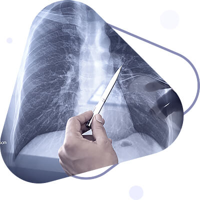
Abscess rupture into the bronchus is accompanied by a large amount of sputum, one or two days before this there may be bad breath, sometimes a small amount of mucopurulent sputum and hemoptysis. The amount of sputum is 250-400 ml, sometimes reaching 1 liter per day. Sputum contains many white blood cells, detritus, fatty acid crystals, Dittrich's plugs, elastic fibers, and a variety of microflora. Most often the sputum is yellow-green in color and, when fasted, gives a characteristic layering: a frothy layer, a purulent layer, and a mucous-serous layer. Radiologically, an isolated solitary cavity filled with fluid and gas, with slight infiltration around it, is defined. The cavity may be large in size.
With good drainage of the cavity and absence of sequesters the abscess may be completely emptied. Its cavity shrinks, the amount of sputum decreases, gradually acquiring a mucous character. General intoxication symptoms decrease, body temperature and blood values normalize. The recovery occurs.
Such character of purulent lung diseases corresponds to the classical idea of lung abscess as a limited accumulation of pus, in which, according to S.I. Spasokokuchenko. Spasokukotsky distinguishes purulent contents, a rounded cavity and a more or less formed wall. A.A. Opokin applied the term "simple abscess" to such processes, and its radiological description was first given by Reader in 1906.
However, a favorable outcome of acute abscess is rare; more often the disease acquires a protracted course, characterized by remissions with periodic delays of sputum discharge. There may be a dissociation between the sputum and the temperature reaction: when the amount of sputum decreases, the body temperature rises (with chills), after its discharge - decreases. Often the sputum becomes stinky and contains an admixture of blood. Sometimes it can be found in the lower crumbly layer (apparently, as a consequence of incomplete purulent melting of pulmonary tissue, indicating the insufficiency of the body's defenses). Radiologically, an increased perifocal reaction zone, sometimes the presence of sequesters in the abscess cavity is determined. On bronchoscopic examination, significant swelling of the mucous membrane of the bronchi and often blockage of their dense purulent secretion is noted.
The outcome of acute abscess depends on the degree of elimination of factors (sequestration, insufficient drainage, etc.), which negatively affect its reverse development and contribute to its prolonged course. The disease in such cases acquires a chronic character.
Chronic abscess most often develops as a result of unfavorable course of acute (or prolonged) lung abscess, and also in cases of festering complication of chronic pneumonia. Changes in the lungs in this case may be of the most diverse nature. There are the following forms of chronic lung abscess: chronic abscess in the form of a single cavity with more or less pronounced fibrous capsule and perifocal zone of pneumonic infiltration; limited pneumosclerosis with multiple, most often of different sizes abscesses; limited piosclerosis of lungs, radiologically manifested as areas of uneven darkening with spots of lumen; limited or spread bronchiectasis.
In accordance with the above, clinical picture of chronic lung abscess is diverse, differing in some cases by poor symptoms (subfebrile temperature, cough with mucopurulent sputum), in others by hemoptysis, large amount of purulent sputum with unpleasant odor, high irregular type of temperature reaction, chills, increased sweating, emaciation, shortness of breath. There is thickening of the distal phalanges of the fingers ("drumsticks") and changes in the fingernails, which look like watch glass.
Various complications are observed:
- septicaemia;
- purulent metastases to the brain;
- repeated pulmonary hemorrhages;
- perforation of abscess into pleural cavity with development of pyopneumothorax.
In some cases amyloidosis develops after long-term course of the disease.
Course. In some cases of acute abscess there is complete recovery, but more often the process becomes chronic and prolonged and fatal from one of complications.
Diagnosing an acute abscess before it has burst into the bronchus can be very difficult. When purulent sputum appears, it can be said that "the diagnosis is written in the spittoon." Recognition of protracted and chronic forms of abscess also causes difficulties in some cases. The most difficult is differential diagnosis when the abscess is localized in the upper parts of the lungs, as well as in those forms that proceed with a small amount of sputum (radiologically they appear as small infiltrates or a limited cavity that does not contain fluid). In such cases, differential diagnosis with tuberculous cavernous disease is based on the following data: perifocal infiltration is more typical for abscessed pneumonia; the abscess cavity is often oval rather than round; specific changes may be detected in tuberculosis around the cavity. Differential diagnostic difficulties may arise in decaying lung cancer and especially in secondary tumor suppuration. In the latter case clinical and radiological picture is similar to that of primary lung abscess. To establish the diagnosis tomographic and bronchoscopic examination, as well as cytological examination of sputum are of great importance. Significant difficulties may arise in differential diagnosis of primary abscess and purulent lung cyst. In the latter case, the process is often recurrent.
As follows from the above, the diagnosis of lung abscess should always be carried out in parallel with finding out its genesis.
Gangrene of the lung (Gangraena pulmonum)
Lung gangrene is the deadening, putrefactive decay of lung tissue, accompanied by foul-smelling sputum. In some cases it occurs as an independent disease (now extremely rare), in others as an outcome of a chronic lung abscess. Sputum of a gangrenous nature may also be detected during exacerbation of a chronic abscess.
At lung gangrene decay of pulmonary tissue has no clearly delineated borders and develops due to significant suppression of the body's defenses.
The main etiological role is played by anaerobic bacteria.
Pathomorphology. On autopsy one or more stinking cavities without distinct borders are found in the lungs. The necrotic area contains cellular detritus and large number of various, mainly anaerobic, bacteria.
Clinic. The disease usually develops acutely, and is characterized by a severe general condition of the patient. A characteristic clinical picture was excellently described by G.I. Sokolsky in the last century: "At the beginning of the disease, unusual pallor of the face, blue circles under the eyes, large exhaustion of energy, small, weak pulse. Soon thereafter the limbs grow cold, the face is covered with sticky sweat, the tongue becomes dry, thirst, delirium, convulsions, death..." And further: "...this dreadful disease is certainly fatal: delirium and hiccups usually precede a quick death. The prescription of ayre root, naphtha, wine and the like continues life only for a short time."
A severe clinical picture, the main symptoms of which are marked intoxication, significant arterial hypotension and the separation of a characteristic stinky sputum, sometimes of chocolate color (containing spirochetes, fusobacteria, putrifying streptococci, etc.), is not observed in all patients and does not always end in death. This was pointed out by Laennec. If acute gangrene is complicated by ichorosis pleurisy, the prognosis is hopeless. In general the clinical picture of pulmonary gangrene is similar to that of acute abscess, but all symptoms are more pronounced, especially intoxication.
There is no self-healing. The mortality rate, according to various authors, used to reach 90%. Recently, due to the use of antibiotics, the prognosis is more favorable.
Bronchiectatic disease (Morbus bronchoectaticus)
Bronchiectatic disease is a special form of suppurative lung disease characterized by bronchiectasis (abnormally dilated bronchi). In this case the bronchi and peribronchial tissue are deeply affected, which is accompanied by the development of destructive changes. Due to joining of infection, purulent process develops, and dilated bronchi are filled with purulent contents.
Etiology and pathogenesis. Most often bronchiectatic disease occurs in connection with pneumonia, often develops against a background of focal or diffuse pneumosclerosis, after infections (measles, whooping cough, tuberculosis, syphilis), as well as after ingress of foreign bodies in bronchi. The mechanism of bronchiectasis development is different in different cases, and therefore we distinguish between retentive, destructive and atelectatic bronchiectasis. Sometimes congenital bronchiectasis is observed, which is usually considered as a malformation - the result of dysplasia of the bronchial wall or interstitial stroma of the lung.

Pathomorphology. A distinction is made between cylindrical, spindle-shaped, and baggy bronchial dilations. Cylindrical and spindle-shaped ones are typical for larger bronchi (more often by their genesis they are retentive), and mesheshotnye - for small ones (usually of destructive origin). The latter cause more severe course of the disease and more frequent disability of patients. There are significant structural changes in bronchial walls - atrophy and death of muscle and elastic fibers, dystrophic changes in cartilage. Due to disturbed drainage function of bronchi, atelectasis, pneumonia, abscesses can develop which subsequently lead to sclerosis of lung segment.
Clinic. If bronchiectases are not subjected to inflammatory changes (not infected), are few in number and small in size, they do not manifest clinically for a long time. The inflammatory (purulent inflammatory) process leads to the development of bronchiectatic disease. In the early period of the disease patients complain of cough with purulent sputum, sometimes with streaks of blood. In cold and wet seasons, patients feel worse (malaise, weakness, increased cough). This condition can last for years. However, often due to repeated acute respiratory diseases, smoking, the process can progress.
The clinical picture in such cases becomes more pronounced:
- increased dyspnea;
- there is more dyspnea, hemoptysis;
- coughing becomes more painful and persistent;
- the amount of sputum increases, which may acquire a foul-smelling character;
- sometimes the sputum may be abundant when the patient takes a certain (for this patient) position;
- general condition worsens;
- periodic increase in body temperature (sometimes in the form of "fever candles");
- the face becomes puffy;
- acrocyanosis, characteristic changes in the distal phalanges of the fingers ("drumsticks");
- emaciation.
Auscultation of the lungs may reveal irregular, "mosaic" breathing (weakened in some parts, rigid in others), as well as dry and moist rales, which are sometimes heard as if at the ear and have a peculiar crackling character. The course is usually prolonged, with periodic exacerbations and various complications: pulmonary bleeding, abscesses, pleural empyema, metastatic abscesses (especially in the brain). The development of pulmonary heart disease and amyloidosis is possible.
Prognosis depends on the severity of the disease, determined by the extent of the process and its clinical course (frequency and severity of exacerbations). In the presence of complications the prognosis is unfavorable. In limited bronchiectasis, especially when localized in one segment or in one lung, when surgical treatment is possible, in some cases recovery is observed.
Diagnosis in cases of a pronounced clinical picture is not very difficult. Of great importance is the presence of purulent sputum and the characteristic radiological picture in tomography and bronchography. The usual radiological examination may reveal intensification of the pulmonary pattern, its honeycomb or cellular nature; sometimes cavities, etc.
Treatment and prophylaxis of purulent lung diseases
Treatment of purulent lung diseases can be carried out both conservatively and surgically. Conservative treatment is effective for acute lung abscess, sometimes for a prolonged course of the disease, and ineffective for chronic forms (in such cases it is either palliative, causing remission of the disease, or used as a preparation for surgical treatment, sometimes in combination with it). The most favorable effect of conservative therapy is observed in cases of lung infections associated with acute pneumonia. The general state and age of the patients are of great importance.
When treating patients with suppurative lung diseases it is necessary to take into account the general changes in the body, disorders of various systems and organs, concomitant diseases, etc.
Treatment should be comprehensive, including the following measures:
- combating infection (prescription of antibiotics, sulfonamide drugs);
- sanation of the bronchial tree and restoration of bronchial patency (mainly intrabronchial administration of various drugs);
- stimulating therapy (blood transfusion, vitamin therapy);
- application of symptomatic means.
In conservative therapy, antibiotics are of primary importance, because in suppressing lung infections is the main task. The following principles of antibiotic therapy should be adhered to:
- prescribe sufficiently high doses of drugs;
- take into account the sensitivity to them of pathogenic microflora of sputum;
- to conduct combined treatment with antibiotics, i.e. to prescribe simultaneously two, rarely three different drugs, periodically alternating them (the technique of sequential alternation of antibiotics according to a cyclic system is particularly important at prolonged and chronic forms of the disease);
- if there is a danger of candida development, prescribe anti-candida antibiotics (nystatin, levorin) and vitamins as early as possible;
- to administer antibiotics in different ways (intravenously, intramuscularly or intravenously, through the respiratory tract, combining different ways of application).
The most effective is the introduction of drugs (bronchodilators, enzymes, antibiotics) through the respiratory tract, which helps to sanitize the bronchi, improve their drainage function and get the drugs closer to the pus. Various methods of administration have been proposed: a) through a rubber catheter inserted into the bronchus (method of bronchial catheterization); b) into the bronchi through a bronchoscope; c) in the form of aerosols. Method of injection through trachea with laryngeal syringe is ineffective.
Segmental or zonal instillation can be performed using bronchoscope (G. I. Burchinsky, B. L. Frantsuzov), which enables to inject antibiotics into bronchi of smaller caliber, corresponding to the segment of purulent focus location and associated with the latter. The technique of segmental instillation is as follows.
Bronchoscopy is performed, the bronchus is cleaned, and after determining the branch leading to the nidus of suppuration and the patency of which is broken, additional anesthesia of its orifice is carried out. Then a thin ureteral catheter is inserted into the lumen of this branch under the control of the eye, and an antibiotic solution is poured into it with an ordinary syringe with a needle placed on it. Penicillin is injected at 300,000-500,000 IU or more, and streptomycin at 500,000 IU every 3-4 days (10-15 injections total).
The main advantages of endobronchial administration of antibiotics are as follows: instillation with a rubber catheter is easily tolerated and indicated in patients even with severe disease; sanation of affected bronchi in the suppuration area is possible. However, resorptive action of antibiotics in this case is low, which requires mandatory simultaneous intramuscular or oral administration. With severe bronchial lesions and widespread process this method is ineffective. Treatment with bronchoscope is the best method of bronchial sanation and restoration of patency of their branches. It is essentially a therapeutic-diagnostic method, allowing to control the dynamics of the process and the efficiency of treatment.
Undoubtedly positive effect is produced by administration of antibiotics in form of aerosols. At the same time there is a pronounced resorptive effect. Aerosol therapy has few contraindications, it is especially effective for widespread bronchial lesions. In limited suppurative processes accompanied by severe bronchial lesions, aerosol therapy is advisable for prevention of bronchogenic dissemination of the process. This method, however, can not replace intrabronchial catheterization, because in severe disorders of drainage bronchial patency the introduction of drugs in the form of aerosol is less effective than bronchial catheterization.
It is advisable to combine different methods of antibiotic administration, which compensates the disadvantages of each of them and summarizes the overall effect.
The method of transthoracic administration of antibiotics in loco, i.e. by puncture through the chest wall (thoracentesis) is also used,
In addition to antibiotics, sulfonamides are used for suppurative lung diseases, especially if there is intolerance to antibiotics, if sputum microflora is insensitive to them, and also if there is a risk of candida development. Combined treatment with antibiotics and sulfonamides is indicated for acute forms of these diseases.
To improve the drainage function of the bronchi and facilitate expectoration of sputum, ephedrine, expectorants (iodine preparations, bromhexine, etc.) are used, especially for viscous sputum, and also use postural (positional) drainage; patients with chronic abscess and bronchiectatic disease are prescribed inhalation of proteolytic enzymes and alkaline solutions.
Stimulating therapy and, first of all, hemotransfusion (blood transfusion by drip method for a long time) plays a significant role in the treatment of suppurative pulmonary diseases. It is especially indicated for pulmonary bleeding, anemia, intoxication, exhaustion, and a sluggish course of the process. Also used are transfusions of plasma, casein hydrolyses, and for the purpose of detoxification therapy - hemodez, polyglucin.
In prolonged infiltrative processes and in the presence of pulmonary tissue sequesters in the abscess cavity UHF-therapy is used. In some cases endobronchial injection of iodolipol is effective. Complications require special therapeutic measures (in most cases - surgery): profuse, recurrent bleeding, pyopneumothorax, pleural empyema.
Persons who have had an acute lung abscess, as well as those suffering from bronchiectatic disease and who have undergone surgery for suppurative lung disease, are subject to dispensary observation. When indicated, they are prescribed a repeated course of conservative treatment.
Prognosis in case of purulent pulmonary disease is conditioned by clinical form of the disease and to a great extent depends on timely and complete treatment measures. At early complex treatment there is a recovery without residual cavity of an abscess, which is extremely important, because even "dry" residual cavity can be a source of process recurrence.
Prevention of lung suppurative diseases consists in full treatment of diseases against which they arise, first of all, pneumonia. People suffering from chronic purulent lung diseases need to be employed, and if the course of the disease is severe - to be transferred to a disability.
By: Dr. Jai Jalaj


